
Devenez membre d'Incathlab et bénéficiez d'un accès complet !

Inscription Connexion
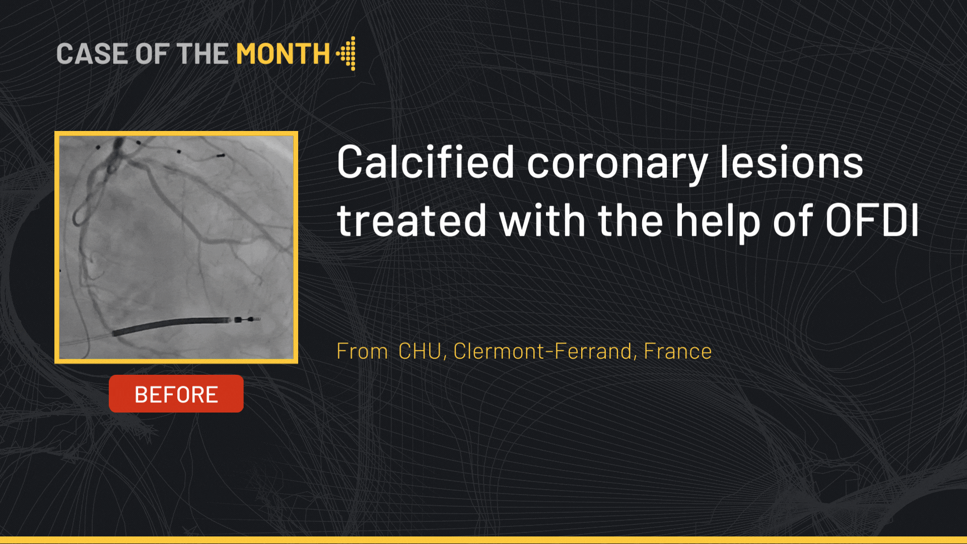
This didactic procedure concerns a 66 years old women presenting bilateral intermittent claudication, Past history: Endarterectomy of left internal carotid artery in 2014.
Global angiography revealed: Firstly, Two Vessels Coronary lesions, Secondly bilateral common femoral severe calcified stenosis, Thirdly Restenosis post endarterectomy of left internal carotid, De novo Right internal carotid stenosis, and occlusion of right brachiocephalic trunk, and occlusion of left subclavian artery.
The first procedure involved recanalization of the subclavian artery with a covered Stent BENTLEY® followed by bilateral Shockwave of Left and Right common femoral arteries and final deployment Supera Stent ®
The Second procedure that we show here consisted of Right intern carotid artery stenting and Brachiocephalic trunk recanalization.
Dual antiplatelet therapy before the procedure and Interruption of anti-hypertensive therapy the day before.
Educational objectives
- How to manage multivascular patient with multiple lesion location: In what order should these different lesions be treated and by which approach?
- Plan a step-by-step carotid artery and brachiocephalic trunk stenting procedure.
- How to manage access through tortuous anatomy? Brachial or Femoral approach?
- How to proceed to a safe and successful catheterization of the right internal carotid artery and brachiocephalic trunk?
- Materials choice: guidewires, protection filter, guiding catheter, balloons and carotid stent.
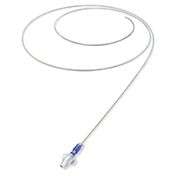
- How to prepare, advance the embolic protection system: FilterWire EZ™ through the lesion and release the filter upstream the lesion?
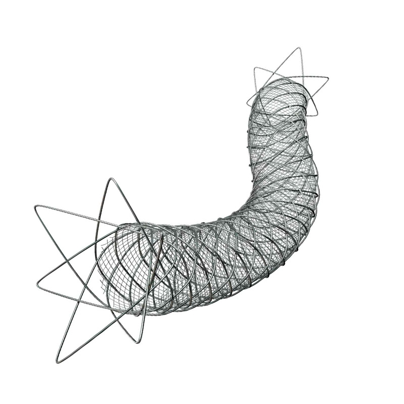
- Tips and tricks for a good positioning and implantation of the Micro Mesh carotid stent.
- How to safely retrieve the embolic protection system: FilterWire EZ™?
- What are adjunctive per procedural pharmacotherapies.
Step-by-step procedure: Right internal carotid artery stenting
1) Access sites:
- Brachial approach : 6 French braided introducer sheath
- Femoral access: 8 French access using micro puncture system.
- Heparin administration.
2) Right common carotid artery catheterization:
- Continuous flushing of the guiding catheter while introducing guidewire, embolic protection device, balloons or stent.
- Advance softly an 6 Fr PENUMBRA Guiding catheter to the subclavian artery over a 0.035’’ Stiff GUIDEWIRE
- Advance the 0.035” Guidewire towards the common carotid artery.
- Switching to 0.035” ADVANTAGE Terumo ® Wire then to AMPLATZ wire in order to have more support
- Advance the selective guiding catheter and the 6 Fr PENUMBRA introducer to the distal part of the common carotid artery with the tip oriented towards the internal carotid ostium.
3) Preparation and deployment of embolic protection system: FilterWire EZ™:
- Preparation of the filter with a special attention to avoid air bubbles.
- Preform the wire tip shape according to the lesion morphology.
- Careful and gentle crossing of the lesion avoiding plaque destabilization.
- Release the filter in a vertical segment of the internal carotid distally to the lesion: be sure to have enough space for stent distal landing zone.
- Verify the good position and the opening of the filter under fluoroscopy.
4) Pre-dilatation
- Atropine administration
- Good balloon preparation: avoid air bubbles to avoid cerebral air embolism in case of balloon rupture.
- Pre dilatation of the lesion using a 4 x 30mm Ultra-Soft™ balloon , inflated to 4 ATM
- Checking pre dilatation result , no more backflow after predilatation
5) Stenting
- Select the precise spot of stent deployment
- Deployment of the ROADSAVER micromesh stent terumo ® 8 x 20 mm
6) Post-dilatation
- Post dilatation of the lesion using a 5 *15 mm Ultra-Soft™ balloon. Inflated to 15 ATM
- Checking post dilatation result.
7) Retrieval of embolic protection system: FilterWire EZ™:
- Check the filter content and the quality of the flow.
- Remove the filter.
- Verify if there is any dissection or spasm.
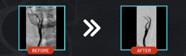
8) Final angiographic control: Cervical and Intra-cranial ( 2 views Frontal & Lateral)
Step-by-step procedure: Brachiocephalic trunk stenting
1) Access sites:
- Brachial approach: 6 French introducer sheath
- Femoral access: 8 French access using micro puncture system.
2) Brachiocephalic trunk artery catheterization:
- Braided Femoral Introducer Sheath 7 French 70 cm crossed gently the Femoral Stent Supera OD ® , and gently placed in the entrance of brachiocephalic trunk
3) Crossing the lesion :
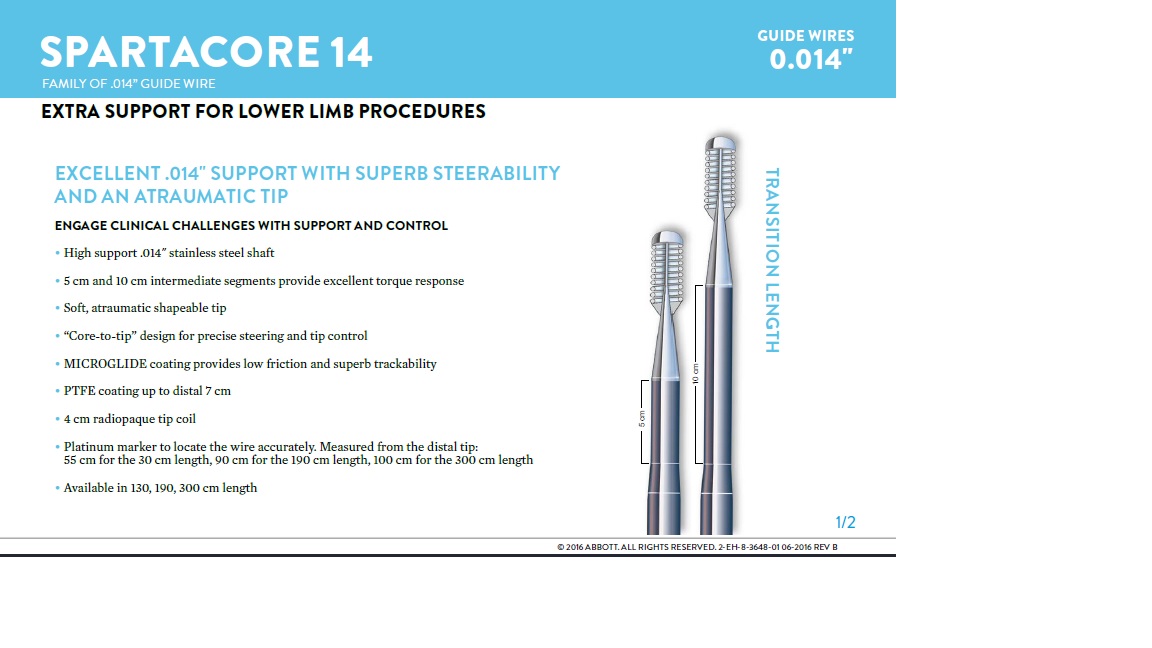
- Using Asahi Halberd® guidewire 0014 via femoral access before switching to Abbott Spartacore ® 0014 guidewire.
- Advance the Abbott Spartacore ® 0014 guidewire towards the common carotid artery in order to have more support.
- Use of the BER II catheter to steer another Abbott Spartacore ® 0014 guide wire to the subclavian artery.
4) Pre-dilatation
- We change to a 0035 guide wire to have more support before predilatation
- Predilatation toward the subclavian artery using 6 x 60 mm Shockwave balloon
5) Stenting brachiocephalic trunk
- Stenting toward the subclavian artery with : Bentley covered stent ® 8X37 mm
- Using caudal view in order to place the stent in the level of the carina between common carotid and the subclavian artery
6) Final angiographic control: Verify if there is any dissection or spasm.
Medical adjunctive treatments
- Pre-procedural: Heparin.
- During procedure : Atropine and ephedrine.
- Post procedural : double therapy: Aspirin 75mg o.d. + Clopidogrel 75mg o.d for one month.
- After one month : Scheduling Coronary PCI.
Bibliography
Dernière mise à jour : 15/01/2024


FilterWire EZ™ / Boston Scientific
Our Cases of the Month
The case of the month is a new way for our users to watch, learn, and share with incathlab. They can watch a video that highlights an innovative case and uses excellent pedagogical techniques, lear...
Suggestions
San Francisco : Mardi 26 mars 2024 de 08h à 09h30 (GMT+1)
New York : Mardi 26 mars 2024 de 11h à 12h30 (GMT+1)
Buenos Aires : Mardi 26 mars 2024 de 13h à 14h30 (GMT+1)
London / Dublin : Mardi 26 mars 2024 de 16h à 17h30 (GMT+1)
Paris / Berlin : Mardi 26 mars 2024 de 17h à 18h30 (GMT+1)
Istanbul : Mardi 26 mars 2024 de 18h à 19h30 (GMT+1)
Moscou / Dubaï : Mardi 26 mars 2024 de 20h à 21h30 (GMT+1)
Bangkok : Mardi 26 mars 2024 de 23h à 00h30 (GMT+1)
Shanghai : Mercredi 27 mars 2024 de 00h à 01h30 (GMT+1)
Tokyo : Mercredi 27 mars 2024 de 01h à 02h30 (GMT+1)
Sydney : Mercredi 27 mars 2024 de 02h à 03h30 (GMT+1)
Wellington : Mercredi 27 mars 2024 de 04h à 05h30 (GMT+1)
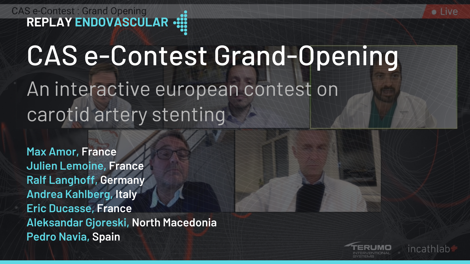
CAS e-Contest Final Results
an interactive european contest on carotid artery stenting
San Francisco : Lundi 10 février 2025 de 07h à 08h (GMT+1)
New York : Lundi 10 février 2025 de 10h à 11h (GMT+1)
Buenos Aires : Lundi 10 février 2025 de 12h à 13h (GMT+1)
London / Dublin : Lundi 10 février 2025 de 15h à 16h (GMT+1)
Paris / Berlin : Lundi 10 février 2025 de 16h à 17h (GMT+1)
Istanbul : Lundi 10 février 2025 de 17h à 18h (GMT+1)
Moscou / Dubaï : Lundi 10 février 2025 de 19h à 20h (GMT+1)
Bangkok : Lundi 10 février 2025 de 22h à 23h (GMT+1)
Shanghai : Lundi 10 février 2025 de 23h à 00h (GMT+1)
Tokyo : Mardi 11 février 2025 de 00h à 01h (GMT+1)
Sydney : Mardi 11 février 2025 de 01h à 02h (GMT+1)
Wellington : Mardi 11 février 2025 de 03h à 04h (GMT+1)
Benefits of braided micromesh
Roadsaver TM Insights Week: Celebrating a Decade of CAS Advances
San Francisco : Mardi 11 février 2025 de 07h à 08h (GMT+1)
New York : Mardi 11 février 2025 de 10h à 11h (GMT+1)
Buenos Aires : Mardi 11 février 2025 de 12h à 13h (GMT+1)
London / Dublin : Mardi 11 février 2025 de 15h à 16h (GMT+1)
Paris / Berlin : Mardi 11 février 2025 de 16h à 17h (GMT+1)
Istanbul : Mardi 11 février 2025 de 17h à 18h (GMT+1)
Moscou / Dubaï : Mardi 11 février 2025 de 19h à 20h (GMT+1)
Bangkok : Mardi 11 février 2025 de 22h à 23h (GMT+1)
Shanghai : Mardi 11 février 2025 de 23h à 00h (GMT+1)
Tokyo : Mercredi 12 février 2025 de 00h à 01h (GMT+1)
Sydney : Mercredi 12 février 2025 de 01h à 02h (GMT+1)
Wellington : Mercredi 12 février 2025 de 03h à 04h (GMT+1)
Radial approach benefits in CAS
Roadsaver TM Insights Week: Celebrating a Decade of CAS Advances
San Francisco : Mercredi 12 février 2025 de 07h à 08h (GMT+1)
New York : Mercredi 12 février 2025 de 10h à 11h (GMT+1)
Buenos Aires : Mercredi 12 février 2025 de 12h à 13h (GMT+1)
London / Dublin : Mercredi 12 février 2025 de 15h à 16h (GMT+1)
Paris / Berlin : Mercredi 12 février 2025 de 16h à 17h (GMT+1)
Istanbul : Mercredi 12 février 2025 de 17h à 18h (GMT+1)
Moscou / Dubaï : Mercredi 12 février 2025 de 19h à 20h (GMT+1)
Bangkok : Mercredi 12 février 2025 de 22h à 23h (GMT+1)
Shanghai : Mercredi 12 février 2025 de 23h à 00h (GMT+1)
Tokyo : Jeudi 13 février 2025 de 00h à 01h (GMT+1)
Sydney : Jeudi 13 février 2025 de 01h à 02h (GMT+1)
Wellington : Jeudi 13 février 2025 de 03h à 04h (GMT+1)
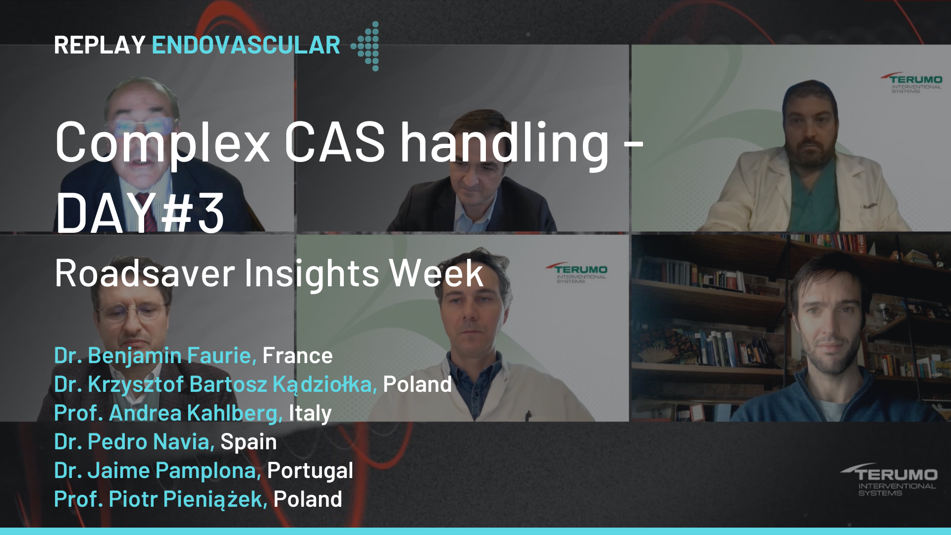
Complex CAS handling
Roadsaver TM Insights Week: Celebrating a Decade of CAS Advances
San Francisco : Jeudi 13 février 2025 de 07h à 08h (GMT+1)
New York : Jeudi 13 février 2025 de 10h à 11h (GMT+1)
Buenos Aires : Jeudi 13 février 2025 de 12h à 13h (GMT+1)
London / Dublin : Jeudi 13 février 2025 de 15h à 16h (GMT+1)
Paris / Berlin : Jeudi 13 février 2025 de 16h à 17h (GMT+1)
Istanbul : Jeudi 13 février 2025 de 17h à 18h (GMT+1)
Moscou / Dubaï : Jeudi 13 février 2025 de 19h à 20h (GMT+1)
Bangkok : Jeudi 13 février 2025 de 22h à 23h (GMT+1)
Shanghai : Jeudi 13 février 2025 de 23h à 00h (GMT+1)
Tokyo : Vendredi 14 février 2025 de 00h à 01h (GMT+1)
Sydney : Vendredi 14 février 2025 de 01h à 02h (GMT+1)
Wellington : Vendredi 14 février 2025 de 03h à 04h (GMT+1)
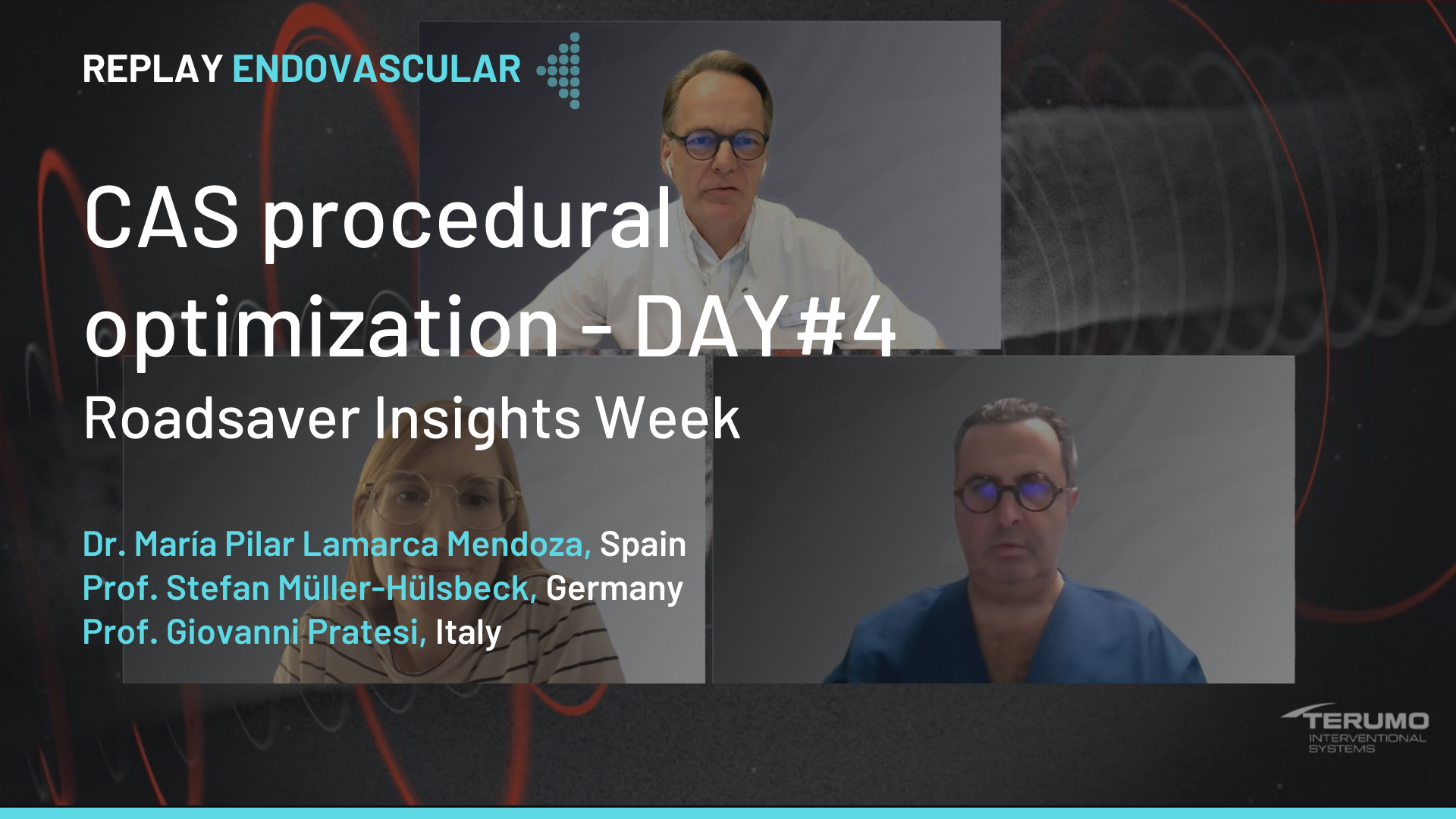
CAS procedural optimization
Roadsaver TM Insights Week: Celebrating a Decade of CAS Advances
San Francisco : Vendredi 14 février 2025 de 07h à 08h (GMT+1)
New York : Vendredi 14 février 2025 de 10h à 11h (GMT+1)
Buenos Aires : Vendredi 14 février 2025 de 12h à 13h (GMT+1)
London / Dublin : Vendredi 14 février 2025 de 15h à 16h (GMT+1)
Paris / Berlin : Vendredi 14 février 2025 de 16h à 17h (GMT+1)
Istanbul : Vendredi 14 février 2025 de 17h à 18h (GMT+1)
Moscou / Dubaï : Vendredi 14 février 2025 de 19h à 20h (GMT+1)
Bangkok : Vendredi 14 février 2025 de 22h à 23h (GMT+1)
Shanghai : Vendredi 14 février 2025 de 23h à 00h (GMT+1)
Tokyo : Samedi 15 février 2025 de 00h à 01h (GMT+1)
Sydney : Samedi 15 février 2025 de 01h à 02h (GMT+1)
Wellington : Samedi 15 février 2025 de 03h à 04h (GMT+1)
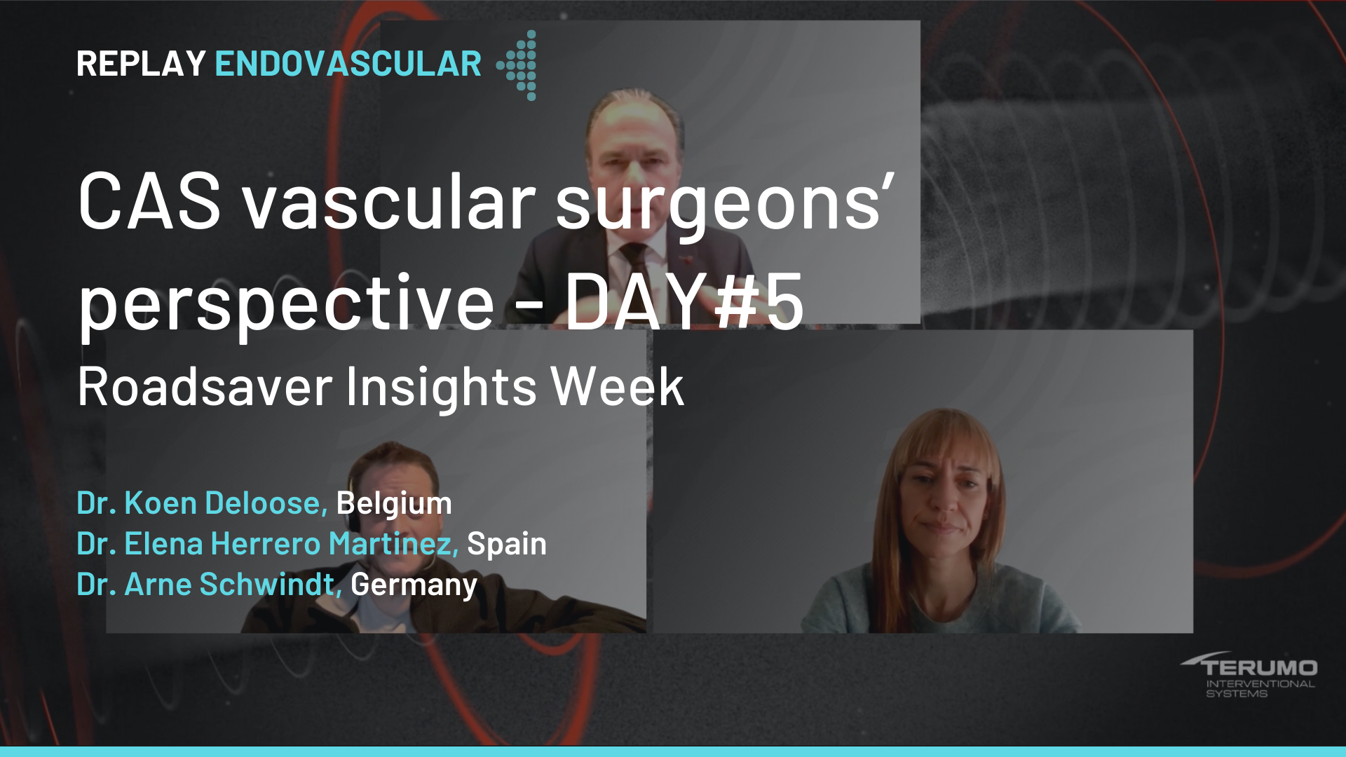
CAS vascular surgeons' perspective
Roadsaver TM Insights Week: Celebrating a Decade of CAS Advances
San Francisco : Jeudi 22 juin 2023 de 08h à 09h30 (GMT+2)
New York : Jeudi 22 juin 2023 de 11h à 12h30 (GMT+2)
Buenos Aires : Jeudi 22 juin 2023 de 12h à 13h30 (GMT+2)
Reykjavik : Jeudi 22 juin 2023 de 15h à 16h30 (GMT+2)
London / Dublin : Jeudi 22 juin 2023 de 16h à 17h30 (GMT+2)
Paris / Berlin : Jeudi 22 juin 2023 de 17h à 18h30 (GMT+2)
Istanbul : Jeudi 22 juin 2023 de 18h à 19h30 (GMT+2)
Moscou / Dubaï : Jeudi 22 juin 2023 de 19h à 20h30 (GMT+2)
Bangkok : Jeudi 22 juin 2023 de 22h à 23h30 (GMT+2)
Shanghai : Jeudi 22 juin 2023 de 23h à 00h30 (GMT+2)
Tokyo : Vendredi 23 juin 2023 de 00h à 01h30 (GMT+2)
Sydney : Vendredi 23 juin 2023 de 02h à 03h30 (GMT+2)
Wellington : Vendredi 23 juin 2023 de 04h à 05h30 (GMT+2)