×
Il semble que vous utilisiez une version obsolète de internet explorer. Internet explorer n'est plus supporté par Microsoft depuis fin 2015. Nous vous invitons à utiliser un navigateur plus récent tel que Firefox, Google Chrome ou Microsoft Edge.
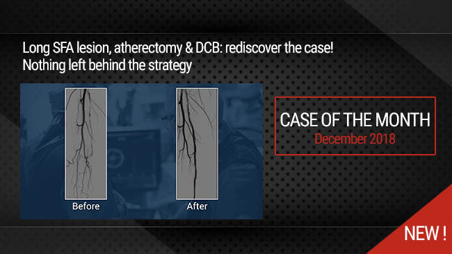
Nothing Left behind Strategy: Long SFA lesion, Directional atherectomy & DCB
Case of the Month: December 2018

This didactic procedure concerns a 62 years old man, presenting severe right limb intermittent claudication (Rutherford 3) & long proximal SFA lesion with subsequent short occlusion.
It was treated by contralateral and retrograde approach, the lesion was prepared with directional atherectomy & a new cutting balloon (Chocolate-Medtronic). A good final result was obtained with DCB angioplasty.
Educational objectives
- How to deal with extensive SFA disease.
- How to performSFA retrograde puncture, crossing and guidewire externalization.
- Effectivness of Directional atherectomy in long SFA lesions.
- Optimal vessel preparation with combined atherectomy & cutting balloon.
- How to leave Nothing behind.
Step-by-Step description
- Left femoral access 7F (Contralateral approach).
- Crossover approach with 6F long sheath assisted by Admiral 6x40mm balloon.
- Approach lesion with 0.018" Command guidewire supported by Trailblazer microcatheter.
- After Command & Connect guidewires failure, a retrograde approach is decided.
- Retrograde puncture of the distal right SFA.
- Retrograde crossing of the lesion & externalization of the command over a BER catheter.
- Filter placment in the distal popliteal artery.
- predilatation with 3.5mm balloon of the occluded zone.
- Directional atherectomy using a HawkOne device 7F.
- Balloon dilatation with a 5x80mm balloon : distal & proximal SFA.
- Second directional atherectomy run.
- Distal SFA dilatation with Chocolate 6x40mm balloon (Medtronic).
- Multilevel dilatation with DCB : IN PACT Pacific 5x120mm (Medtronic)
- Distal filter retreiving Spider 7mm (Medtronic).
- Final angiographic control.
Protocol
- Procedure time: 90 min
- Exposure time: 29 min
- Exposure: 321 mGy
-
Contrast volume: 120 ml
Biobliography
-
-
Combined HawkOne directional atherectomy and paclitaxel-coated balloon angioplasty for isolated calcified popliteal artery lesion: a no-stent approach to lower extremity endovascular revascularization. - Article
Authors: Loffroy R, Chevallier O, Falvo N, Gehin S, Midulla M, Galland C.
Publication:doi: 10.21037/qims.2018.03.10.
-
Directional Atherectomy Followed by a Paclitaxel-Coated Balloon to Inhibit Restenosis and Maintain Vessel Patency: Twelve-Month Results of the DEFINITIVE AR Study. - Article
Authors: Zeller T, Langhoff R, Rocha-Singh KJ
Publication doi:10.1161/CIRCINTERVENTIONS.116.004848.
-
Debulking Atherectomy in the Peripheral Arteries: Is There a Role and What is the Evidence? - Artice
Authors: Katsanos K, Spiliopoulos S, Reppas L, Karnabatidis D.
Publication doi: 10.1007/s00270-017-1649-6. Epub 2017 Apr 27.
-
Combined treatment of heavy calcified femoro-popliteal lesions using directional atherectomy and a paclitaxel coated balloon: One-year single centre clinical results. - Article
Authors: Cioppa A, Stabile E, Popusoi G, Salemme L, Cota L
Publication doi: 10.1016/j.carrev.2012.04.007. Epub 2012 May 25.
-
A prospective, multi-center study of the chocolate balloon in femoropopliteal peripheral artery disease: The Chocolate BAR registry. - Article
Authors: Mustapha JA, Lansky A, Shishehbor M, Miles McClure J, Johnson S, Davis T, Makam P, Crowder W, Konstantino E, Attaran RR; Chocolate Bar Investigators.
Publication doi: 10.1002/ccd.27565. Epub 2018 Mar 7.
Vidéos liées
Nothing Left behind Strategy: Long SFA lesion, Directional atherectomy & DCB
Case of the month: December 2018
Partager
Playlists liées

Our Cases of the Month
The case of the month is a new way for our users to watch, learn, and share with incathlab. They can watch a video that highlights an innovative case and uses excellent pedagogical techniques, lear...
Partager