Today’s Standard for Modern Endovascular Intervention: See, prep, and treat
By: Bianca Mulaney-Topkar, MD, MS MPH

At the 2025 Cardiovascular and Interventional Radiological Society of Europe (CIRSE) conference, the session “Today’s standard for modern endovascular intervention: see, prep, and treat”, sponsored by Boston Scientific, brought together global experts in peripheral artery disease (PAD) to review the latest in endovascular revascularization techniques. Moderated by Dr Marianne Brodmann (Medical University of Graz, Austria), the discussion centered on vessel preparation, drug-eluting therapies, atherectomy, and intravascular ultrasound. Below is a summary of each of the four presentations with key takeaways.
PRESENTATION 1: Definitive treatment? Today’s evidence.
Gunnar Tepe, MD, Head of Diagnostic and Interventional Radiology, Klinikum Rosenheim, Germany
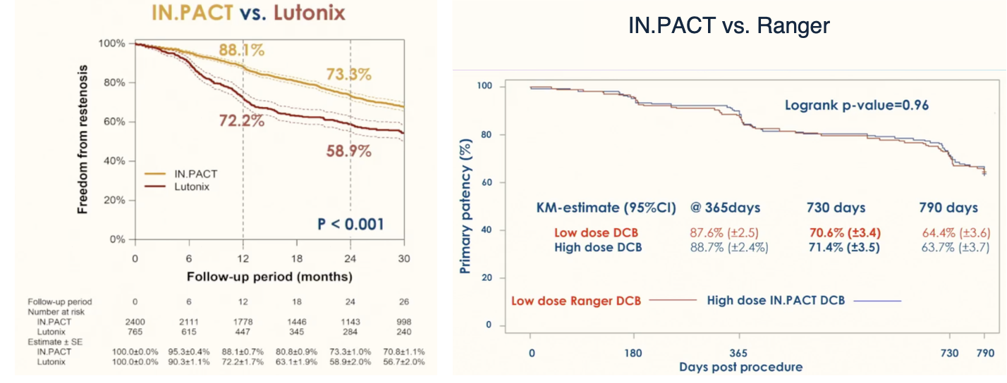
Dr. Tepe highlighted a shift in practice over recent years to drug-eluting therapy for femoropopliteal lesions, given the significantly higher patency compared to plain balloon angioplasty and bare metal stenting. However, not all drug-coated balloons have the same efficacy. For example, IN.PACT and Ranger balloons have similar patency, but IN.PACT balloons have superior patency to Lutonix (88% vs. 72% freedom from restenosis at 12 months, p<0.001).1,2
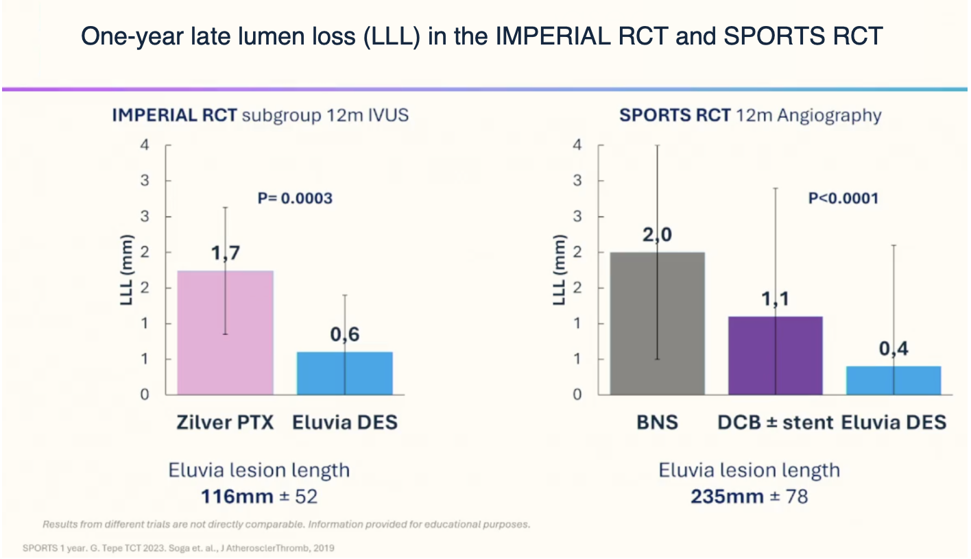
In addition, the rate of rescue stenting following drug-coated balloon angioplasty (DCBA) due to complications such as dissection remains high, especially in longer lesions. For example, 58% of DCBA treatments required a bail-out stent in the SPORTS trial.3 For this reason Dr. Tepe advocated for drug-eluting stenting (DES) as first-line for long (i.e., >15-20cm length) superficial femoral artery (SFA) lesions. Data from the IMPERIAL RCT4,5 as well as a forthcoming meta-analysis6 and the SPORTS RCT3 demonstrate both primary and secondary outcomes (including vessel diameter stenosis/lumen loss at 12 months, primary patency at 1 and 2 years, and freedom from target lesion revascularization at 2 years) were best in the paclitaxel eluting stent (DES) group compared to paclitaxel-coated balloon (DCB) and bare nitinol stent (BNS) in long-segment SFA (TASC C/D) lesions.
Key takeaway: For short (e.g., mid/distal SFA) or more diffuse lesions, drug-coated balloons are ideal. For long SFA lesions, drug-eluting stenting is superior to both bare metal stenting and drug-coated balloons.
PRESENTATION 2: What lies beneath? The utility of IVUS.
Daniel van den Heuvel, MD, Interventional Radiologist, St Antonius Hospital, Nieuwegein, Netherlands
Angiography remains the workhorse imaging modality for endovascular treatment of femoropopliteal disease. Intravascular ultrasound (IVUS) is known to be expensive – so what is the utility of adding IVUS to conventional angiography? IVUS allows for better visualization of plaque morphology and lesion length, leading to more accurate sizing of interventions and a “leave nothing behind” approach, when compared to conventional angiography alone.
|
Phase |
Utility of IVUS |
|
Pre-intervention |
Determine plaque morphology and severity |
|
Intra-procedural |
Size vessel diameter for device, ensure location of crossing wire is intraluminal |
|
Post-intervention |
Evaluate success of treatment (residual stenosis, look for dissection, whether additional post-dilation/stenting needed) |
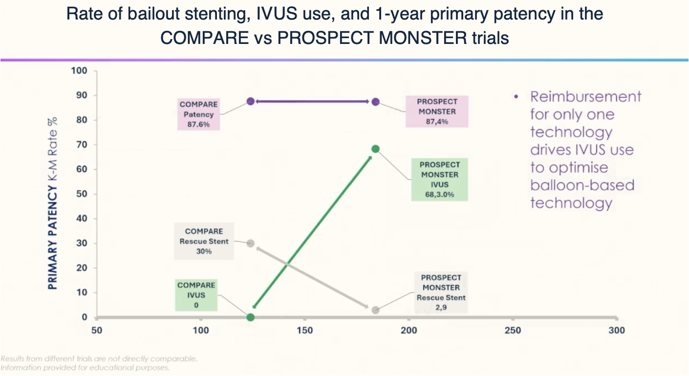
The rate of intraprocedural bailout stenting was significantly lower in the PROSPECT MONSTER trial (2-3%)7 versus the COMPARE trial (26-30%).8 Why? Although other differences between the trials may exist, Dr. van den Heuvel notes that the rate of IVUS-guided DCB therapy was significantly higher in PROSPECT-MONSTER (68.3%) than in COMPARE (0%), suggesting that the use of IVUS was responsible for the decrease in bailout stenting.
Similarly, Ko et al. found that the use of IVUS to guide DCBA of femoropopliteal lesions resulted in larger balloon size (5mm vs 4.5mm), higher inflation pressure (11.8mmHg vs 8.9mmHg), and higher rate of adjuvant post-dilation (31% vs 16%), ultimately leading to higher primary patency (86.6% vs 74.8%) and freedom from clinically-driven target lesion revascularization (92.4% vs 83.0%) at one year compared to angiography alone.9 This was especially the case for more complex TASC C/D lesions.10 Follow-up studies show that the benefit of decreased restenosis with IVUS persisted at 2 and 3 years.11,12
As Dr. van den Heuvel explained, although formal randomized control trials and cost-effectiveness studies of IVUS versus angiography do not exist, IVUS substantially decreases the need for reinterventions (estimated at €22,000)13 and bail-out stenting, which may justify the cost of IVUS in clinical practice. Experts seem to agree that IVUS is “worth it” for above-the-knee interventions, especially in patients with claudication, where there may be more of a question whether to stent (whereas it may not make as much of a difference in patients with CLTI where the stent rate is higher). Regarding below-the-knee lesions, the TIBIA RCT is currently underway and will hopefully have preliminary results soon.
Key takeaway: with conventional angiography, we may be more likely to undersize or not treat the full extent of lesions. IVUS enables more accurate sizing and treatment of femoropopliteal lesions, decreasing the need for reinterventions and bail-out stenting.
PRESENTATION 3: Minimize stenting? – The utility of atherectomy.
Athanasios Diamantopoulos, MD, PhD, Consultant Interventional Radiologist, Guy’s and St Thomas' Hospital, London, UK
Nearly half (42%) of infrainguinal lesions are chronic total occlusions (CTOs)14, and we know that heavy calcification significantly worsens DCB efficacy. For example, while the studies cited in presentation 1 noted >80% primary patency at 1 year following DCBA, lesions with circumferential 270-360° calcification have 50% primary patency at 1 year.15 Atherectomy is particularly useful for vessel preparation in heavily calcified lesions as it debulks plaque, improves vessel compliance, and increases drug transfer to tissues when followed by drug-eluting therapy. Of note, atherectomy devices have a wide variety of underlying mechanisms (e.g., laser, orbital, directional, rotational) and we will not delve into comparisons here—Dr. Diamantopoulos focused on Boston Scientific’s Jetstream atherectomy device, which uses front cutting, rotational blades to engage chronically calcified lesions with active aspiration to minimize embolization risk. In particular, three studies demonstrate the outcomes of Jetstream atherectomy with DCBA:
1. The J-SUPREME trials showed that Jetstream with DCBA was superior to Jetstream with plain balloon angioplasty, with 97% vs 40% primary patency at 6 months, and 3.4% vs 26% target lesion revascularization at 12 months.16
2. The Ellipse prospective multicenter observational study demonstrated a 12-month freedom from target lesion revascularization of 96% following Jetstream with DCBA (no comparison group).17
3. The CORVUS trial showed that Jetstream with DCBA had similar primary patency and freedom from target lesion revascularization when compared to stenting alone, though rates of distal embolization were higher with atherectomy.18
Regarding the risks of distal embolization, Dr. Diamantopoulos shared some practical tips: 1) treat only long occlusions with atherectomy, 2) first treat the middle segment of the lesion, then slowly treat the proximal cap, then finally treat the distal cap, and 3) after atherectomy is complete, place a filter for embolic protection and do DCBA over the filter to minimize the risks.Ultimately, Dr. Diamantopoulos advocates that atherectomy can effectively downgrade TASC complexity and enable a DCB-only approach. Especially in complex TASC C/D lesions, atherectomy can help maintain greater than 90% primary patency with a low (2-7%) rescue stent rate, as shown in the JET FORWARD and JETSTREAM LL registries.
Key takeaway: atherectomy’s value may lie in debulking and vessel prep for long, heavily calcified lesions like CTOs. By effectively downgrading lesion complexity, atherectomy reduces the need for rescue stents and supports DCB-only strategies even in longer lesions.
PRESENTATION 4: Better patient outcomes? – Getting it right the first time.
Yan Gouëffic, MD, PhD, Professor of Vascular Surgery, Groupe Hospitalier Paris Saint Joseph, Paris, France
Dr. Gouëffic highlighted the importance of good vessel preparation for long-term patency. Greater than 30% residual stenosis at the time of DCBA, was associated with worse primary patency and need for reintervention at 1 year. 19 Similarly, residual spiral dissection at the time of DCBA was associated with worse primary patency (69.8% vs 83.3%) and freedom from target lesion revascularization (78.7% vs 93%) at 1 year.20 Given this, he argues that we should rethink our strategy after initial balloon angioplasty – if there is concern for residual stenosis or dissection, strongly consider switching from DCBA to drug-eluting stenting (DES). Why not plain stenting? Well, the primary patency of DES is superior to non-DES at 1 year in this pooled meta-analysis (unpublished data):
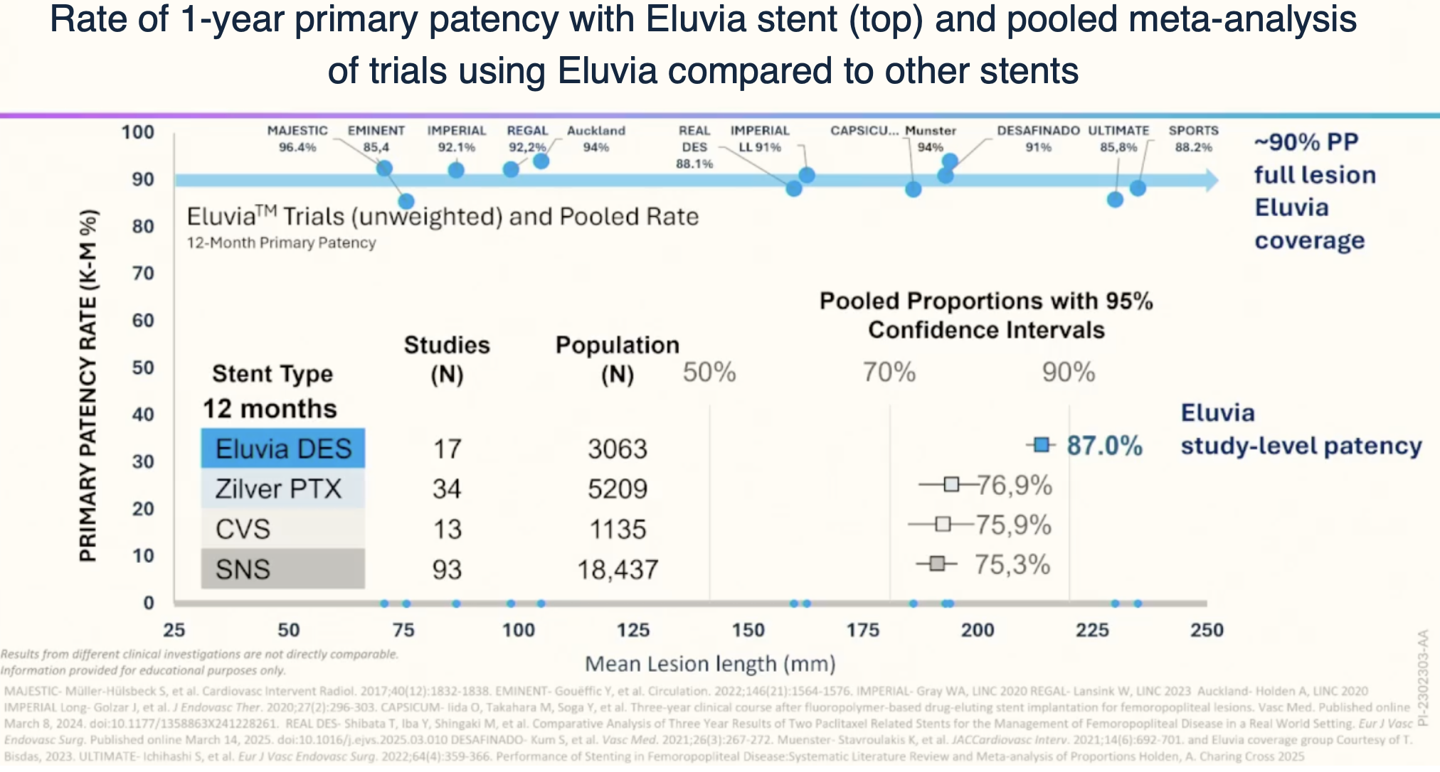
Dr. Gouëffic also noted that drug-coated stents such as Zilver PTX exhibit a decline in patency as lesion length increases, whereas drug-eluting stents such as Eluvia maintain patency even with long lesions.
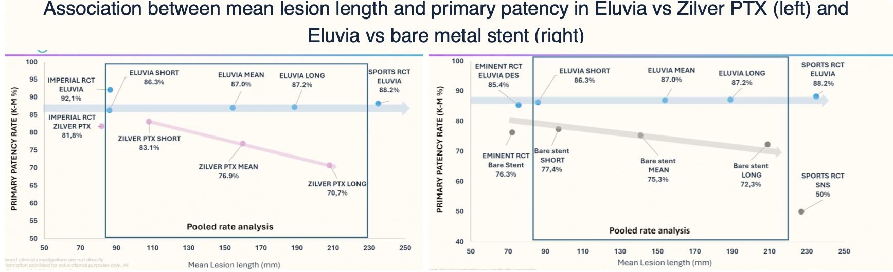
Key takeaway: Scaffolding, ideally with drug-eluting stenting, is recommended in lesions with >30% residual stenosis or dissection after initial vessel prep, >10 cm lesion length, and CTOs.
Conclusion
Drug-eluting therapies have emerged as the gold standard for femoropopliteal disease. For short lesions, DCB is often adequate. IVUS improves lesion assessment and intervention sizing while reducing bailout stenting and reintervention rates. For heavily calcified lesions and CTOs, atherectomy can debulk and optimize vessel compliance, enhancing drug uptake when used in conjunction with DCB. While atherectomy may reduce the need for stenting, it has a higher risk of distal embolization. For long, calcified lesions, or situations where there is residual stenosis or dissection after balloon angioplasty, drug-eluting stents are recommended.
About the author
Bianca Mulaney-Topkar, MD, MS, MPH is an integrated vascular surgery resident at Massachusetts General Hospital in Boston, MA and winner of the 2025 Drug Eluting Technology (DET) Show hosted by Incathlab. She is also a Marshall Scholar and incoming National Clinician Scholars Program (NCSP) fellowship recipient.
Moderator and panelist biographies
Dr Marianne Brodmann is the Head of Division of Angiology, Head of Clinical Research at Division of Angiology at the Medical University of Graz, Austria.
Dr Brodmann is a Fellow of the European Society of Cardiology, Board Member of the European Union of Medical Specialist (UEMS), past President of the ÖGA (Austrian Society of Angiology) and National Delegate of the IUA (International Union of Angiology). Research activities include anticoagulation and thrombolysis, new endovascular technologies in the peripheral field and evaluation of the nature of restenosis. She also founded a research establishment for experimental angiology animal models at the University of Graz. Dr Brodmann has participated in over 50 international clinical trials including Principal Investigator for the Bioflex I, Lutonix BTK and TOBA-BTK clinical trials and Steering Committee of the CVI Ingenuity trial.
Gunnar Tepe, MD is Professor of Radiology, Head of Diagnostic and Interventional Radiology at the Academic Hospital RoMed Clinic of Rosenheim in Rosenheim, Germany.
He started his medical career at the University of Tuebingen. In 2003 he was elected as professor of Radiology. He led the interventional section in the department of radiology at the University of Tuebingen for more than 5 years and started several clinical studies in that time which he also led as a PI. One of his landmark trials was the Thunder study. It was the first human usage of drug-coated-balloons in patients with peripheral arterial disease. The data of that study were published in the New England Journal of Medicine. In 2009 Prof. Tepe moved to the RoMed Clinic Rosenheim and became the head of the Department of Diagnostic and Interventional Radiology. From 2009 to 2019 he was a part of the organizing committee of Angio Update and since 2017 Prof. Tepe is a member of the Charing Cross Executive Board. Disclosure(s): BBraun, Boston Scientific: Grant/Research Support (Ongoing) He has disclosed that he is a paid consultant to and receives grant/research funding from Cook Medical, Medrad, Inc., and Medtronic, Inc. Dr. Tepe may be reached at [email protected].
Daniel van den Heuvel, MD is an Interventional Radiologist at the Saint Antonius Hospital
in Nieuwegein, The Netherlands. He earned his doctorate of medicine from the University of Amsterdam in 2001. After a two-year residency at the Department of Vascular Surgery he started his training as a resident Radiology in the St. Antonius Hospital in 2003 and was a fellow at the department of Interventional Radiology from 2009 until 2011. His specialty is peripheral artery disease with a focus on CLTI including venous arterialization. In addition to this he is specialized in the treatment of pulmonary arteriovenous malformations. He is involved in the training of residents in general Radiology and is program director for residents Interventional Radiology in their 3rd – 5th year.
Dr Athanasios (Thanos) Diamantopoulos, MD, PhD is a Consultant Interventional
Radiologist at Guy’s and St Thomas' and a Senior Clinical Lecturer (Hon) at King’s College London. He qualified in 2004 from the medical school of University of Patras in Greece. He holds a PhD in experimental prevention of contrast induced nephropathy. He has been trained in all aspects of general and interventional radiology and he has completed a fellowship at Guy’s and St Thomas prior to being appointed as a consultant at the same hospital. He has been board certified by the European Board of Interventional Radiologists (EBIR) and is the current clinical lead for Interventional Radiology at Guys and St Thomas Interventional Radiology Department.
Dr Diamantopoulos has authored or co-authored many peer-reviewed original scientific papers and book chapters. In addition, he has given many scientific presentations and is regularly invited as a lecturer in National and International conferences. He is the primary investigator (PI) and co-investigator in numerous studies and has been awarded a Fellowship of the Cardiovascular and Interventional Radiology Society of Europe (FCIRSE) for his contribution in interventional radiology and cardiovascular imaging. Since 2019 he serves as an elected member in the research committee of CIRSE. He specialises in minimally invasive, image guided vascular, non-vascular and interventional oncology procedures both in adults and children. He distinguishes himself by performing cutting-edge procedures that include all aspects of basic and advanced interventional radiology procedures. In an effort to deliver the best possible results, Dr Diamantopoulos likes to incorporate the latest technological advances into his treatment methods such as drug eluting technologies, carbon dioxide angiography, the latest atherectomy techniques and all the newest ablation technologies.
Yann Gouëffic, MD, PhD, is a professor of vascular surgery in the Vascular Center at Groupe Hospitalier Paris Saint Joseph, Paris, France.
He is also President of the Association of the French University Hospitals for Clinical Research. Pr Gouëffic received his MD and PhD from the Nantes university and completed his education from Paris university hospitals and Hope Heart Institute (Seattle, USA). Pr Gouëffic is the former chief of department of vascular surgery at university hospital at Nantes, France, from 2011-2019. Pr Gouëffic performs endovascular and open procedures for peripheral arterial diseases with more than 20 years of experience in this field. His clinical interests include lower extremity arterial disease, endovascular revascularization, wound healing and amputation prevention.
Pr Gouëffic performs a series of studies in these areas and conducts numerous clinical trials. He is the author of numerous peer-reviewed original and review articles, reviewer of different journals. He has consulted on the regulatory pathway and pivotal trials for many vascular devices, has participated in numerous meetings with the French National Authority for Health (Haute Autorité de santé - HAS) and has testified at HAS and FDA panel hearings.
Pr Gouëffic is member of the French Society of Vascular and Endovascular Surgery. He is a co-director of the Paris Vascular Insights meeting. He serves as the editor of the L’Interventionnel magazine. He can be reached at [email protected].
Research funding from Predisurge, Biotronik, WL Gore, GE Healthcare Honoraria from Abbott, BD, Bentely, Biotronik, Boston Scientific, Cook, Eclevar Medtech, GE Healthcare, Ivascular, Medtronic, Penumbra, Sensome, Shockwave medical, WL Gore (consulting, medical advisory board, educational course, speaking)
Vidéos liées

San Francisco : Vendredi 4 avril 2025 de 06h à 08h (GMT+2)
New York : Vendredi 4 avril 2025 de 09h à 11h (GMT+2)
Buenos Aires : Vendredi 4 avril 2025 de 10h à 12h (GMT+2)
Reykjavik : Vendredi 4 avril 2025 de 13h à 15h (GMT+2)
London / Dublin : Vendredi 4 avril 2025 de 14h à 16h (GMT+2)
Paris / Berlin : Vendredi 4 avril 2025 de 15h à 17h (GMT+2)
Istanbul : Vendredi 4 avril 2025 de 16h à 18h (GMT+2)
Moscou / Dubaï : Vendredi 4 avril 2025 de 17h à 19h (GMT+2)
Bangkok : Vendredi 4 avril 2025 de 20h à 22h (GMT+2)
Shanghai : Vendredi 4 avril 2025 de 21h à 23h (GMT+2)
Tokyo : Vendredi 4 avril 2025 de 22h à 00h (GMT+2)
Sydney : Samedi 5 avril 2025 de 00h à 02h (GMT+2)
Wellington : Samedi 5 avril 2025 de 02h à 04h (GMT+2)
The Drug Eluting Technology Show
Playlists liées
THE DET SHOW 2025
This playlist includes 4 video extracts from the DET SHOW 2025: - Live case procedure from UK - Restenosis challenge - Drug-Eluting Technologies in PAD: DES and DCB vs BMS and PO...
