×
Il semble que vous utilisiez une version obsolète de internet explorer. Internet explorer n'est plus supporté par Microsoft depuis fin 2015. Nous vous invitons à utiliser un navigateur plus récent tel que Firefox, Google Chrome ou Microsoft Edge.

Devenez membre d'Incathlab et bénéficiez d'un accès complet !

Vous devez être membre pour accéder aux vidéos Incathlab sans limitation. Inscrivez vous gratuitement en moins d'une minute et accédez à tous les services Incathlab ! Vous avez aussi la possibilité de vous connecter directement avec votre compte facebook ou twitter en cliquant sur login en haut à droite du site.
Inscription Connexion
Inscription Connexion
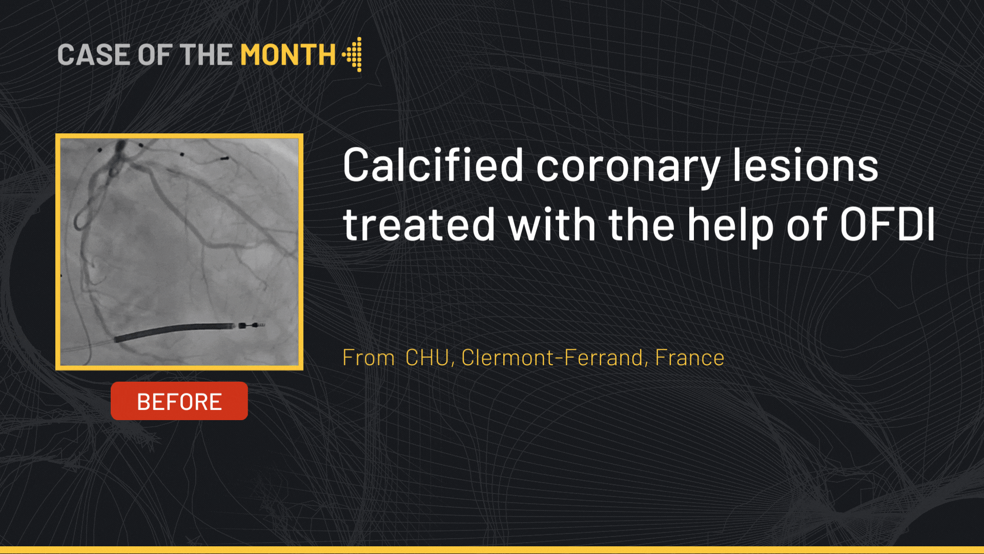
This didactic procedure concerns a 81 years old man presenting with symptomatic severe calcified and ulcerated left carotid on echography. Angiography revealed a long-ulcerated stenosis at the origin of the left internal carotid artery.
Educational objectives
- Plan a step-by-step carotid artery stenting procedure.
- How to manage access through tortuous anatomy?
- How to proceed to a safe and successful catheterization of the left carotid artery?
- Materials choice: guidewires, filter, guiding catheter, balloons and stent.
- How to prepare, advance the embolic protection system: FilterWire EZ™ through the lesion and release the filter upstream the lesion?
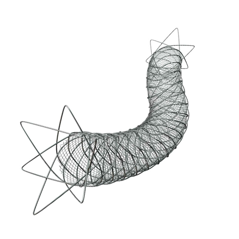
- Tips and tricks for a good positioning and implantation of the Micromesh Roadsaver® stent
- How to safely retrieve the embolic protection system: FilterWire EZ™?
- What are adjunctive per procedural pharmacotherapies?
Step-by-step procedure: Left internal carotid artery stenting
1) Access sites:
- Femoral access: Echo guided 8 French access using micro puncture system.
- Heparin administration.
2) Left common carotid artery catheterization: coaxial technique
- Continuous purging of the guiding catheter while introducing guidewire, embolic protection device, balloons or stent.
- Advance an 8 Fr Hockey Stick guiding catheter to the aortic arch on a 0.035’’ GLIDEWIRE ADVANTAGE® Guidewire.
- Gentle Catheterization of the ostium of the left common carotid artery.
- Advance the 0.035” ADVANTAGE® Guidewire to the external carotid artery.
- Advance a Beacon® Tip 5.0 Fr catheter on the 0.035” ADVANTAGE® Guidewire.
- Exchange to a 0.035” Amplatz Super Stiff™ Guidewire to provide enough support to the Hockey stick 8 Fr guiding catheter to navigate through tortuosity using the coaxial system.
- Advance the guiding catheter to the distal part of the common carotid artery with the tip oriented towards the internal carotid ostium.
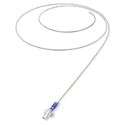
3) Preparation and deployment of embolic protection system: FilterWire EZ™:
- Preparation of the filter with a special attention to avoid air bubbles.
- Preform the wire tip shape according to the lesion morphology.
- Careful and gentle crossing of the lesion avoiding plaque destabilization.
- Release the filter in a vertical segment of the internal carotid distally to the lesion: be sure to have enough space for stent distal landing zone.
- Verify the good position and the opening of the filter under fluoroscopy.
4) Pre-dilatation
- Atropine administration
- Good balloon preparation: avoid air bubbles to avoid cerebral air embolism in case of balloon rupture.li>
- Pre dilatation of the lesion using a 5.5*20mm Ultra-Soft™ balloon.
- Checking pre dilatation result
5) Stenting
- Select the precise spot of stent deployment
- Deployment of the Roadsaver 9*20mm stent.
- Be sure that the dual layer markers are on either side of the lesion.
6) Post-dilatation
- Post dilatation of the lesion using a 6*20mm Ultra-Soft™ balloon.
- Checking post dilatation result.
7) Retrievement of embolic protection system: FilterWire EZ™:
- Check the filter content and the quality of the flow.
- Remove the filter.
- Verify if there is any dissection or spasm.
8) Final angiographic control: Cervical and Intra-cranial
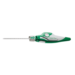
9) Vascular femoral closure with an 8 Fr Angio-Seal™
10) Medical adjunctive treatments
- Pre-procedural: Heparin and Antibioprophylaxis.
- Post procedural: Triple therapy: Aspirin 75mg o.d. + Clopidogrel 75mg o.d. + Enoxaparin 100 UI/kg b.i.d. : 15 days.
- DUS 15 days after.
- Stop Clopidogrel and continue Aspirin 75mg b.i.d and NAOC.
Bibliography
Procedure
- Procedure time: 25 min
- Exposure time: 20,4 min
- R: 296 mgy
- Contrast volume : 120ml
Date du tournage : 27/08/2020
Dernière mise à jour : 11/07/2023
Dernière mise à jour : 11/07/2023


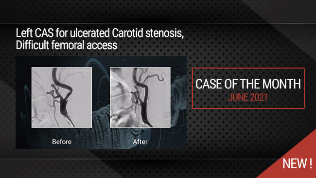

Our Cases of the Month
The case of the month is a new way for our users to watch, learn, and share with incathlab. They can watch a video that highlights an innovative case and uses excellent pedagogical techniques, lear...
Partager
Participer à la discussion
Suggestions
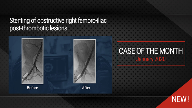
Stenting of obstructive right femoro-iliac post-thrombotic lesions
Case of the month: January 2020
Partager
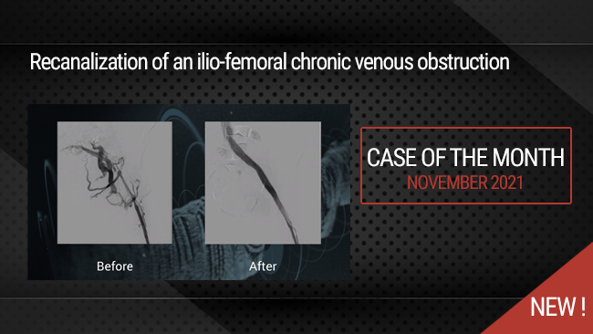
Recanalization of an ilio-femoral chronic venous obstruction
Case of the month: November 2021
Partager
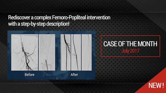
Long Femoral Occlusion (35 cm) - Subintimal crossing and extra long stenting
Case of the month: July 2017
Partager
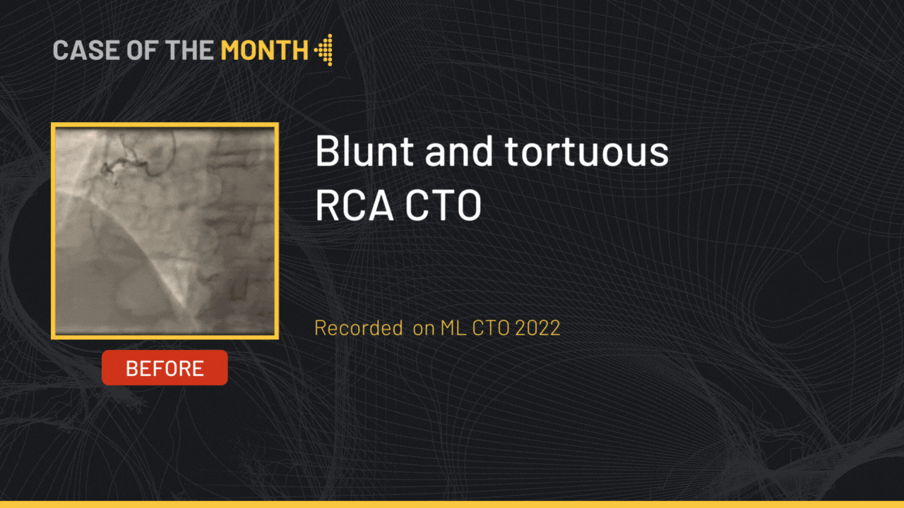
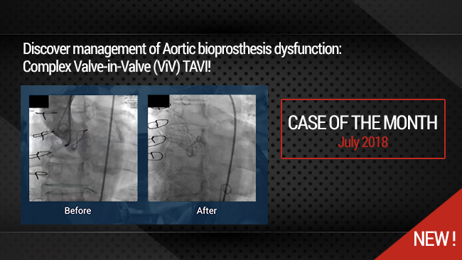
Complex Valve-in-Valve (VIV) TAVI for Aortic bioprosthesis dysfunction
Case of the month: July 2018
Partager
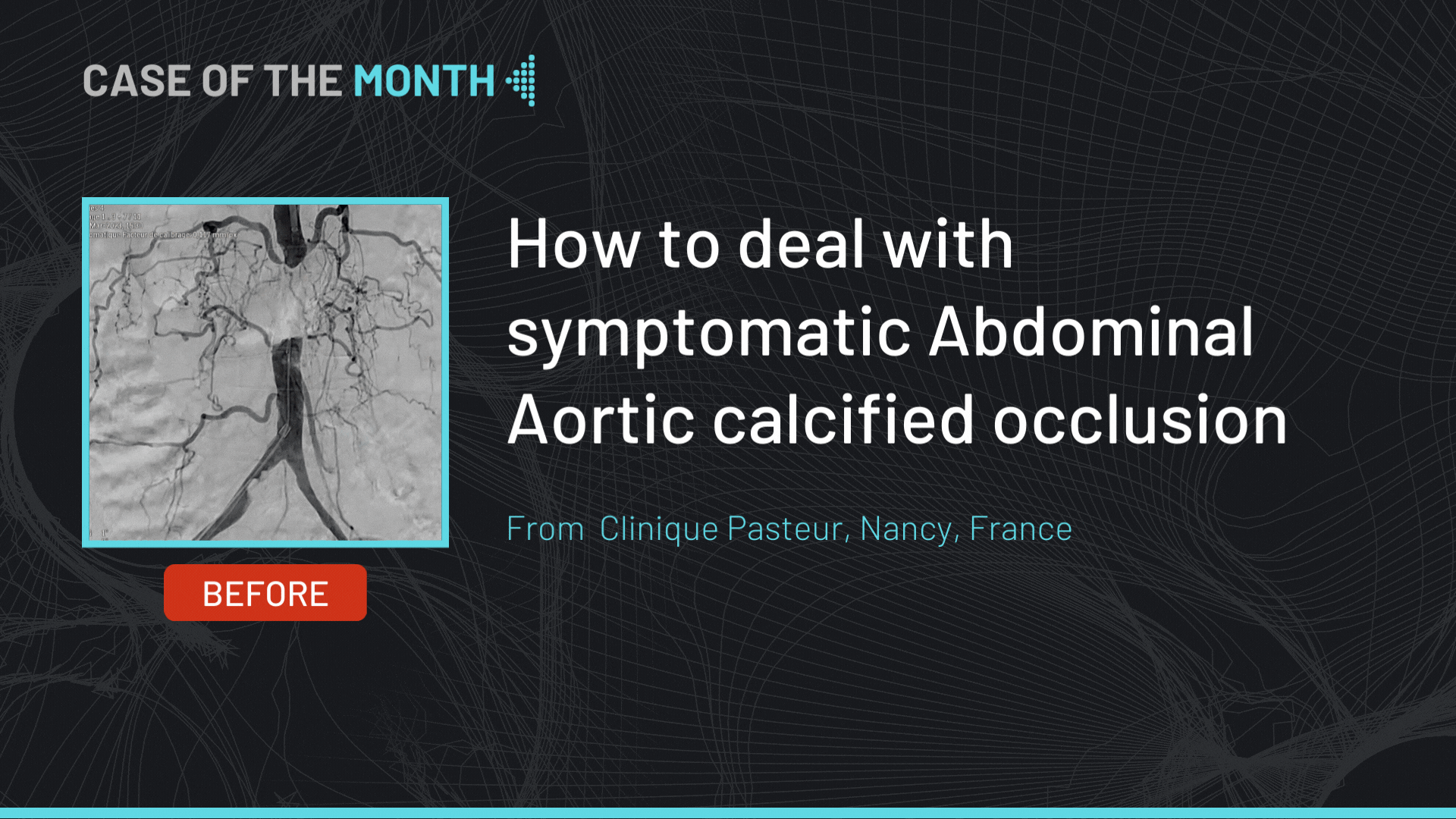
How to deal with symptomatic Abdominal Aortic calcified occlusion
Case of the month: May 2023
Partager
Endres J. To push the amplatz guidwire into the carotid bifurcation is a hazardous maneuver!
aksüyek A. is triple therapy routine in your practice?
Milan M. Its a 50% Stenosis!