
Become an Incathlab member and receive full access to its content!

Registration Login

Left Internal Carotid Artery Occlusion with Progressive Right Internal Carotid Artery Stenosis
This 18 minutes didactic recorded procedure concerns a 68 years male with coronary insufficiency and carotid lesions. He presents an occluded left internal carotid artery associated with a severe ulcerated worsening right internal carotid stenosis.
This complex stenosis was treated by new micromesh carotid stent under carotid protection by filter . Watch this informative procedure on how to treat patients with multiple carotid lesions.
Step-by-Step Procedure
-
Femoral access for carotid angioplasty
-
Common carotid access in tortuous aorta
-
Analysis of carotid lesion by selective angiography
-
Placement of filter as embolic protecting device
-
Pre-dilatation and artery preparation before carotid artery stenting
-
Placement of braided micromesh stent Roadsaver
-
Post Stenting dilatation and analysis of results
-
Filter retrieval and evaluation of carotid circulation
Learning points
-
How to access from groin the brachio-cephalic trunk in a tortuous aorta
-
The use of two guidewires to access common carotid
-
Placement of filter in a tortuous carotid artery
-
Predilatation and preparation of carotid lesion before stenting
-
Accurate placement of new micromesh carotid stent
-
Cardiac monitoring during carotid stenting
Bibliography
-
Mesh-covered (Roadsaver) stent as a new treatment modality for symptomatic or high-risk carotid stenosis - Article
Machnik R, Paluszek P, Tekieli Ł, Dzierwa K, Maciejewski D, Trystuła M, Brzychczy A, Banaszkiewicz K, Musiał R, Pieniążek P.
Postepy Kardiol Interwencyjnej. 2017;13(2):130-134. doi: 10.5114/pwki.2017.68139. Epub 2017 May 30.
-
Acute Occlusions of Dual-Layer Carotid Stents After Endovascular Emergency Treatment of Tandem Lesions - Article
Yilmaz U, Körner H, Mühl-Benninghaus R, Simgen A, Kraus C, Walter S, Behnke S, Faßbender K, Reith W, Unger MM.
Stroke. 2017 Aug;48(8):2171-2175. doi: 10.1161/STROKEAHA.116.015965. Epub 2017 Jul 5.
-
The Casper carotid artery stent: a unique all metal micromesh stent designed to prevent embolic release - Article
Diaz O, Lopez G, Roehm JOF Jr, De la Rosa G, Orozco F, Almeida R.
J Neurointerv Surg. 2017 Apr 24. pii: neurintsurg-2016-012913. doi: 10.1136/neurintsurg-2016-012913.
-
The CLEAR-ROAD study: evaluation of a new dual layer micromesh stent system for the carotid artery - Article
Bosiers M, Deloose K, Torsello G, Scheinert D, Maene L, Peeters P, Müller-Hülsbeck S, Sievert H, Langhoff R, Bosiers M, Setacci C.
EuroIntervention. 2016 Aug 5;12(5):e671-6. doi: 10.4244/EIJY16M05_04.
-
Safety of Slender 5Fr Transradial Approach for Carotid Artery Stenting With a Novel Nitinol Double-Layer Micromesh Stent - Article
Kedev S, Petkoska D, Zafirovska B, Vasilev I, Bertrand OF.
Am J Cardiol. 2015 Sep 15;116(6):977-81. doi: 10.1016/j.amjcard.2015.05.063. Epub 2015 Jun 25.
-
Initial clinical experience with the micromesh Roadsaver carotid artery stent for the treatment of patients with symptomatic carotid artery disease - Article
Hopf-Jensen S, Marques L, Preiß M, Müller-Hülsbeck S.
J Endovasc Ther. 2015 Apr;22(2):220-5. doi: 10.1177/1526602815576337.
-
One swallow does not a summer make but many swallows do: accumulating clinical evidence for nearly-eliminated peri-procedural and 30-day complications with mesh-covered stents transforms the carotid revascularisation field - Article
Musiałek P, Hopkins LN, Siddiqui AH.
Postepy Kardiol Interwencyjnej. 2017;13(2):95-106. doi: 10.5114/pwki.2017.69012. Epub 2017 Jul 19.
Last update : 2021-05-11

OptiRAY® / Guerbet
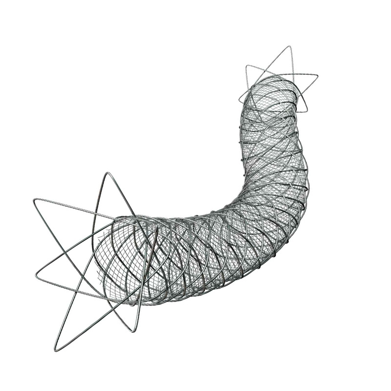

FilterWire EZ™ / Boston Scientific

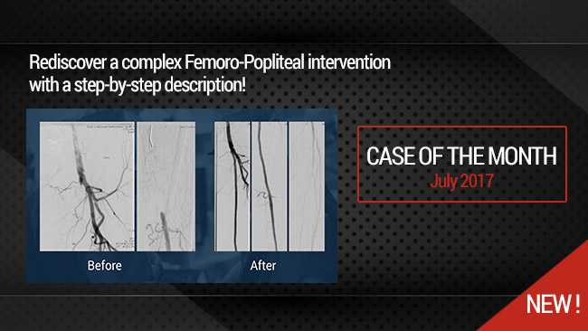
Our Cases of the Month
The case of the month is a new way for our users to watch, learn, and share with incathlab. They can watch a video that highlights an innovative case and uses excellent pedagogical techniques, lear...
Workshop on Complex PCI (Clinique Louis Pasteur - Nancy)
We are pleased to announce you that the Guerbet Masterclass which took place on September 2017 at the Clinique Louis Pasteur (Nancy, France) is available online for all participants. Rediscove...

arie B. 'ישא 'שד איק ןמגןבשאןםמ שדטצפאםצשאןב פשאןקמא 'ןאי ךקדד איקמ 50% דאקמםדןד?.
Max A. dont understand . Sorry
AWADHESH D. Nice
Max A. Thank U
Zambonialbe A. Very interesting case !
Alexander P. super
Max A. Thank you
Chun-Yuan C. Will direct stenting be another choice ?
Max A. Sorry for the delay .
I recommend to predilate for this micromesh Stent to be sure to have an harmonious deployment to easen the crossing . It is particularly important with the CGuard stent
Milan M. Nice example! According to your experience, how do the Micromash Stents behave in highly calcified lesions? Thank You
Max A. In very calcified lesions it is indispensable to prepare the lesion by a pre-dilatation in order to be sure that the residual stenosis is not Severe .
Mohammed R. Good
Max A. Thank You
Maher J. Perfect job
Maher J. Nice job