×
It looks like you're using an obsolete version of internet explorer. Internet explorer is no longer supported by Microsoft since the end of 2015. We invite you to use a newer browser such as Firefox, Google Chrome or Microsoft Edge.

Become an Incathlab member and receive full access to its content!

You must be an Incathlab member to access videos without any restrictions. Register for free in one minute and access all services provided by Incathlab.You will also be able to log into Incathlab from your Facebook or twitter account by clicking on login on the top-right corner of Incathlab website.
Registration Login
Registration Login
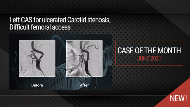
This didactic procedure concerns a 81 years old man presenting with symptomatic severe calcified and ulcerated left carotid on echography. Angiography revealed a long-ulcerated stenosis at the origin of the left internal carotid artery.
Educational objectives
- Plan a step-by-step carotid artery stenting procedure.
- How to manage access through tortuous anatomy?
- How to proceed to a safe and successful catheterization of the left carotid artery?
- Materials choice: guidewires, filter, guiding catheter, balloons and stent.
- How to prepare, advance the embolic protection system: FilterWire EZ™ through the lesion and release the filter upstream the lesion?
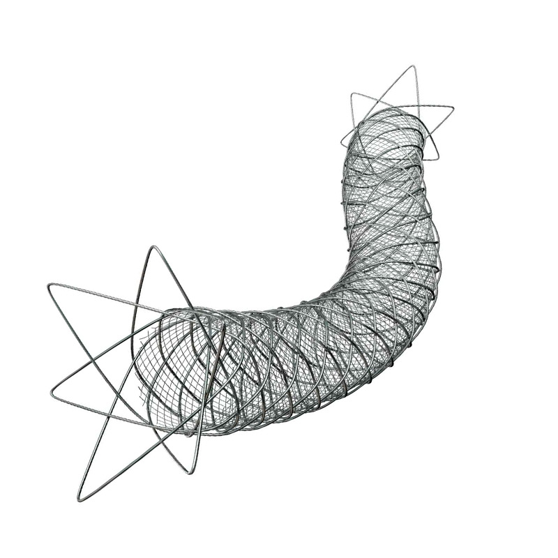
- Tips and tricks for a good positioning and implantation of the Micromesh Roadsaver® stent
- How to safely retrieve the embolic protection system: FilterWire EZ™?
- What are adjunctive per procedural pharmacotherapies?
Step-by-step procedure: Left internal carotid artery stenting
1) Access sites:
- Femoral access: Echo guided 8 French access using micro puncture system.
- Heparin administration.
2) Left common carotid artery catheterization: coaxial technique
- Continuous purging of the guiding catheter while introducing guidewire, embolic protection device, balloons or stent.
- Advance an 8 Fr Hockey Stick guiding catheter to the aortic arch on a 0.035’’ GLIDEWIRE ADVANTAGE® Guidewire.
- Gentle Catheterization of the ostium of the left common carotid artery.
- Advance the 0.035” ADVANTAGE® Guidewire to the external carotid artery.
- Advance a Beacon® Tip 5.0 Fr catheter on the 0.035” ADVANTAGE® Guidewire.
- Exchange to a 0.035” Amplatz Super Stiff™ Guidewire to provide enough support to the Hockey stick 8 Fr guiding catheter to navigate through tortuosity using the coaxial system.
- Advance the guiding catheter to the distal part of the common carotid artery with the tip oriented towards the internal carotid ostium.
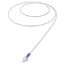
3) Preparation and deployment of embolic protection system: FilterWire EZ™:
- Preparation of the filter with a special attention to avoid air bubbles.
- Preform the wire tip shape according to the lesion morphology.
- Careful and gentle crossing of the lesion avoiding plaque destabilization.
- Release the filter in a vertical segment of the internal carotid distally to the lesion: be sure to have enough space for stent distal landing zone.
- Verify the good position and the opening of the filter under fluoroscopy.
4) Pre-dilatation
- Atropine administration
- Good balloon preparation: avoid air bubbles to avoid cerebral air embolism in case of balloon rupture.li>
- Pre dilatation of the lesion using a 5.5*20mm Ultra-Soft™ balloon.
- Checking pre dilatation result
5) Stenting
- Select the precise spot of stent deployment
- Deployment of the Roadsaver 9*20mm stent.
- Be sure that the dual layer markers are on either side of the lesion.
6) Post-dilatation
- Post dilatation of the lesion using a 6*20mm Ultra-Soft™ balloon.
- Checking post dilatation result.
7) Retrievement of embolic protection system: FilterWire EZ™:
- Check the filter content and the quality of the flow.
- Remove the filter.
- Verify if there is any dissection or spasm.
8) Final angiographic control: Cervical and Intra-cranial
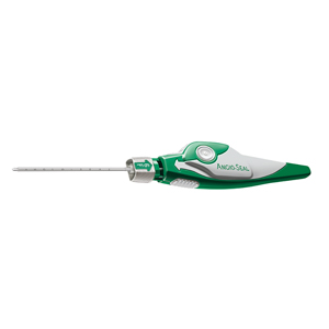
9) Vascular femoral closure with an 8 Fr Angio-Seal™
10) Medical adjunctive treatments
- Pre-procedural: Heparin and Antibioprophylaxis.
- Post procedural: Triple therapy: Aspirin 75mg o.d. + Clopidogrel 75mg o.d. + Enoxaparin 100 UI/kg b.i.d. : 15 days.
- DUS 15 days after.
- Stop Clopidogrel and continue Aspirin 75mg b.i.d and NAOC.
Bibliography
Procedure
- Procedure time: 25 min
- Exposure time: 20,4 min
- R: 296 mgy
- Contrast volume : 120ml
Shooting date : 2020-08-27
Last update : 2023-07-11
Last update : 2023-07-11


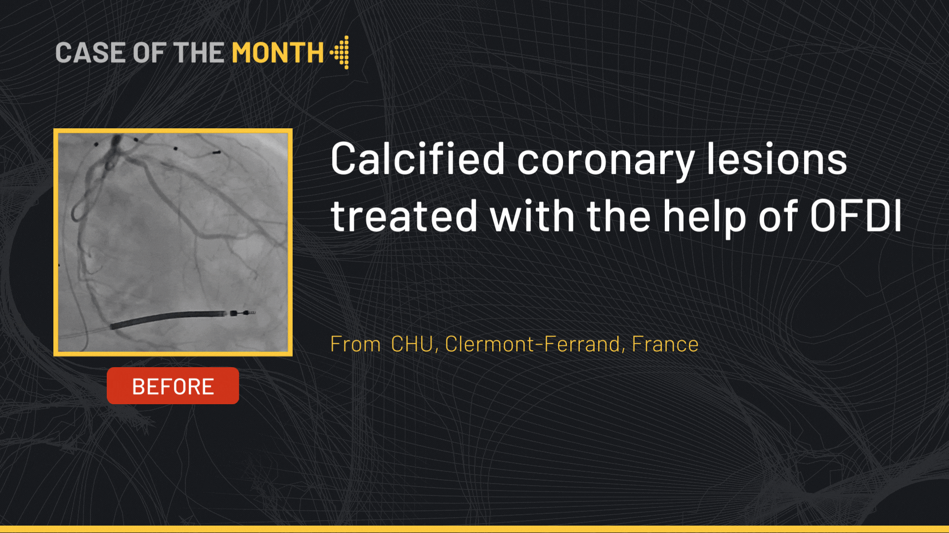
Our Cases of the Month
The case of the month is a new way for our users to watch, learn, and share with incathlab. They can watch a video that highlights an innovative case and uses excellent pedagogical techniques, lear...
Share
Join the Discussion
Suggestions
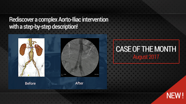
Percutaneous EndoVascular Aortic Repair (PEVAR) with low profile endovascular graft
Case of the month: August 2017
Share
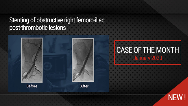
Stenting of obstructive right femoro-iliac post-thrombotic lesions
Case of the month: January 2020
Share
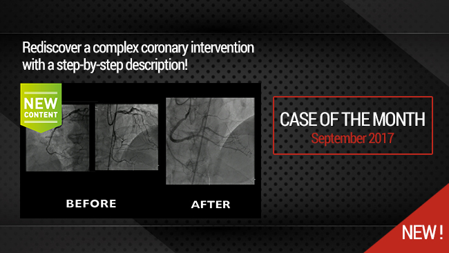
Very complex Mid RCA occlusion
Retrograde in 1st intention and Antegrade approach for recanalization
Share
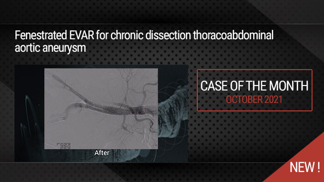
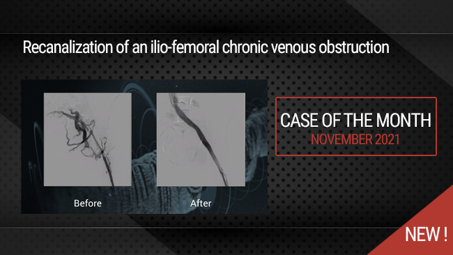
IMAC 2019 : Fenestrated EVAR for chronic dissection thoracoabdominal aortic aneurysm
Case of the month: October 2021
Share
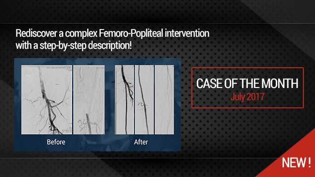
Long Femoral Occlusion (35 cm) - Subintimal crossing and extra long stenting
Case of the month: July 2017
Share
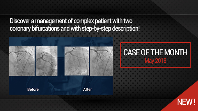
Simultaneous treatment of two coronary artery bifurcations in three vessels disease patient
Dedicated coronary bifurcation stents - Case of the month: May 2018
Share
Endres J. To push the amplatz guidwire into the carotid bifurcation is a hazardous maneuver!
aksüyek A. is triple therapy routine in your practice?
Milan M. Its a 50% Stenosis!